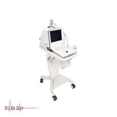
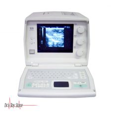
Toshiba Nemio 10 Ultrasound Machine
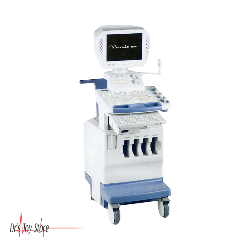
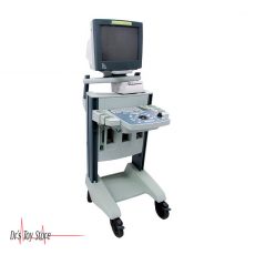
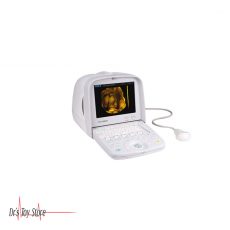
The Toshiba Nemio 10 Ultrasound Machine is a flexible, compact system what delivers superb grayscale image quality, facilitated by the same digital beamformer technology available on all of the Nemio systems. Its ergonomic design provides simplicity and efficiency. And with its highly scalable platform, your diagnostic capabilities can be enhanced to conform to your expanding clinical needs.
Toshiba Nemio 10 Ultrasound Machine Features
- Touch customizable console
- Programmable pop-up menus, screen layout and interface
- 16 user-defined presets
- User-programmable Measurements
- Convex and linear transducer capability
- Two active transducer ports, plus two resting ports
- Read and write zoom
- Cine memory
- Floppy disk drive
- Fully digital continuous beamformer
Meet the new standard in compact, high-performance ultrasound from the leader, Toshiba. Designed and engineered to meet the real-world requirements of busy hospitals and clinics, Toshiba Nemio 10 Ultrasound Machine has ease of use and versatility gives you the ultimate in sophisticated ultrasound diagnostic imaging in a stylish, compact unit. That’s why they call it the Premium Compact.
Toshiba Nemio 10 Ultrasound Machine Applications
- OB/GYN
- Small Parts
High-contrast Resolution – Wide system dynamic range improves high-contrast resolution. Furthermore, the Tissue Harmonic Imaging (THI) technique provides vivid and sharp images
Good Color Sensitivity – 4-way Color technology permits fine and sharp observation of blood flow and allows accurate measurement of blood-flow waveforms
Fusion 3D – Toshiba’s Fusion 3D, which provides 3-dimensional representations of B-mode cross-sectional images of tissues in combination with Power Angio images, is highly useful for tumor observation from all angles and for determination of the treatment.
Panoramic View – Continuous ultrasound images acquired through linear movement of the transducer can be reconstructed into a single wide-field image for display
QSP / PSP – Quad Signal Processing (QSP) / Parallel Signal Processing (PSP) allows receipt of echoes from up to four directions in response to a single transmission pulse. This technique allows diagnosis with higher frame rates.
TDI-Quantification – Tissue Doppler Imaging (TDI) for color display of myocardial dynamics and TDI-Quantification for quantitative evaluation of the results are both provided. Together, these two functions permit full-scale cardiovascular examinations.


Reviews
There are no reviews yet.